Mostrar el registro sencillo del ítem
Detection and removal of dust artifacts in retinal images via sparse-based inpainting
| dc.contributor.author | Barrios, Erik | |
| dc.contributor.author | Sierra, Enrique | |
| dc.contributor.author | Romero, Lenny A. | |
| dc.contributor.author | Millán, María S. | |
| dc.contributor.author | Marrugo Hernández, Andrés Guillermo | |
| dc.date.accessioned | 2022-01-27T14:53:16Z | |
| dc.date.available | 2022-01-27T14:53:16Z | |
| dc.date.issued | 2021-09-06 | |
| dc.date.submitted | 2022-01-26 | |
| dc.identifier.citation | Erik Barrios, Enrique Sierra, Lenny A. Romero, María S. Millán, Andres G. Marrugo. Opt. Pura Apl. 54 (3) 1-14 (2021). DOI: 10.7149/OPA.54.3.51060 | spa |
| dc.identifier.uri | https://hdl.handle.net/20.500.12585/10414 | |
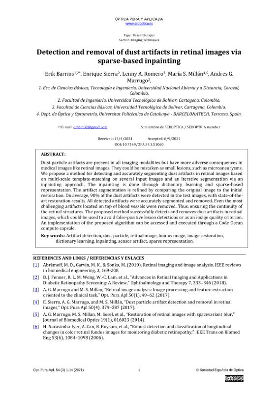
| dc.description.abstract | Dust particle artifacts are present in all imaging modalities but have more adverse consequences in medical images like retinal images. They could be mistaken as small lesions, such as microaneurysms. We propose a method for detecting and accurately segmenting dust artifacts in retinal images based on multi-scale template-matching on several input images and an iterative segmentation via an inpainting approach. The inpainting is done through dictionary learning and sparse-based representation. The artifact segmentation is refined by comparing the original image to the initial restoration. On average, 90% of the dust artifacts were detected in the test images, with state-of-theart restoration results. All detected artifacts were accurately segmented and removed. Even the most challenging artifacts located on top of blood vessels were removed. Thus, ensuring the continuity of the retinal structures. The proposed method successfully detects and removes dust artifacts in retinal images, which could be used to avoid false-positive lesion detections or as an image quality criterion. An implementation of the proposed algorithm can be accessed and executed through a Code Ocean compute capsule | spa |
| dc.format.extent | 14 Páginas | |
| dc.format.mimetype | application/pdf | spa |
| dc.language.iso | eng | spa |
| dc.rights.uri | http://creativecommons.org/licenses/by-nc-nd/4.0/ | * |
| dc.source | Óptica pura y aplicada - vol. 54 n° 3 (2021) | spa |
| dc.title | Detection and removal of dust artifacts in retinal images via sparse-based inpainting | spa |
| dcterms.bibliographicCitation | Abràmoff, M. D., Garvin, M. K., & Sonka, M. (2010). Retinal imaging and image analysis. IEEE reviews in biomedical engineering, 3, 169-208. | spa |
| dcterms.bibliographicCitation | B. J. Fenner, R. L. M. Wong, W.-C. Lam, et al., “Advances in Retinal Imaging and Applications in Diabetic Retinopathy Screening: A Review,” Ophthalmology and Therapy 7, 333–346 (2018) | spa |
| dcterms.bibliographicCitation | A. G. Marrugo and M. S. Millan, “Retinal image analysis: Image processing and feature extraction oriented to the clinical task,” Opt. Pura Apl 50(1), 49–62 (2017). | spa |
| dcterms.bibliographicCitation | E. Sierra, A. G. Marrugo, and M. S. Millán, “Dust particle artifact detection and removal in retinal images,” Opt. Pura Apl 50(4), 379–387 (2017) | spa |
| dcterms.bibliographicCitation | A. G. Marrugo, M. S. Millan, M. Sorel, et al., “Restoration of retinal images with spacevariant blur,” Journal of Biomedical Optics 19(1), 016023 (2014). | spa |
| dcterms.bibliographicCitation | H. Narasimha-Iyer, A. Can, B. Roysam, et al., “Robust detection and classification of longitudinal changes in color retinal fundus images for monitoring diabetic retinopathy,” IEEE Trans on Biomed Eng 53(6), 1084–1098 (2006). | spa |
| dcterms.bibliographicCitation | P. F. Ordóñez, C. M. Cepeda, J. Garrido, et al., “Classification of images based on small local features: a case applied to microaneurysms in fundus retina images,” Journal of Medical Imaging 4(4), 041309 (2017). | spa |
| dcterms.bibliographicCitation | A. Manjaramkar and M. Kokare, “Statistical geometrical features for microaneurysm detection,” Journal of digital imaging 31(2), 224–234 (2018). | spa |
| dcterms.bibliographicCitation | M. Zamfir, E. Steinberg, Y. Prilutsky, et al., “Image defect map creation using batches of digital images,” (2010). Patent US 2010/0321537 A1. | spa |
| dcterms.bibliographicCitation | R. G. Willson, M. W. Maimone, A. E. Johnson, et al., “An optical model for image artifacts produced by dust particles on lenses,” 8th International Symposium on Artificial Intelligence, Robotics, and Automation in Space (i-SAIRAS) (2005). | spa |
| dcterms.bibliographicCitation | C. Li, K. Zhou, and S. Lin, “Removal of dust artifacts in focal stack image sequences. ,” in Proceedings of the 21st International Conference on Pattern Recognition (ICPR2012), 2602–2605, IEEE (2012). | spa |
| dcterms.bibliographicCitation | H. Altamar-Mercado, A. Patino-Vanegas, and A. G. Marrugo, “Extended Focused Image in White Light Scanning Interference Microscopy,” in Imaging and Applied Optics 2019, ITh1C.3, Optical Society of America, (Munich) (2019). | spa |
| dcterms.bibliographicCitation | C. Zhou and S. Lin, “Removal of image artifacts due to sensor dust,” in Computer Vision and Pattern Recognition, 2007. CVPR’07. IEEE Conference on, 1–8, IEEE (2007) | spa |
| dcterms.bibliographicCitation | A. D. Mora, J. Soares, and J. M. Fonseca, “A template matching technique for artifacts detection in retinal images,” in 2013 8th International Symposium on Image and Signal Processing and Analysis (ISPA), 717–722, IEEE (2013). [doi:10.1109/ispa.2013.6703831] | spa |
| dcterms.bibliographicCitation | M. Niemeijer, M. D. Abramoff, and B. van Ginneken, “Image structure clustering for image quality verification of color retina images in diabetic retinopathy screening,” Medical image analysis 10(6), 888–898 (2006). [doi:10.1016/j.media.2006.09.006]. | spa |
| dcterms.bibliographicCitation | M. S. Millan, A. G. Marrugo, and F. Alba-Bueno, “Quality Changes in Fundus Images of Pseudophakic Eyes,” Opt. Pura Apl. 51(4), 50015:1–8 (2018). | spa |
| dcterms.bibliographicCitation | T. Köhler, A. Budai, M. F. Kraus, et al., “Automatic no-reference quality assessment for retinal fundus images using vessel segmentation,” in Proceedings of the 26th IEEE International Symposium on Computer-Based Medical Systems, 95–100, IEEE (2013). | spa |
| dcterms.bibliographicCitation | D. Veiga, C. Pereira, M. Ferreira, et al., “Quality evaluation of digital fundus images through combined measures.,” Journal of Medical Imaging 1(1), 014001 (2014). | spa |
| dcterms.bibliographicCitation | S. A. A. Shah, A. Laude, I. Faye, et al., “Automated microaneurysm detection in diabetic retinopathy using curvelet transform,” Journal of Biomedical Optics 21(10), 101404 (2016). | spa |
| dcterms.bibliographicCitation | P. Yang, L. Chen, J. Tian, et al., “Dust particle detection in surveillance video using salient visual descriptors,” Computers & Electrical Engineering 62, 224–231 (2017). | spa |
| dcterms.bibliographicCitation | L. Chen, D. Zhu, J. Tian, et al., “Dust particle detection in traffic surveillance video using motion singularity analysis,” Digital Signal Processing 58, 127–133 (2016). | spa |
| dcterms.bibliographicCitation | L. Hu, L. Chen, and J. Cheng, “Gray spot detection in surveillance video using convolutional neural network,” in 2018 13th IEEE Conference on Industrial Electronics and Applications (ICIEA), 2806– 2810, IEEE (2018). [doi:10.1109/ICIEA.2018.8398187]. | spa |
| dcterms.bibliographicCitation | D. A. Forsyth and J. Ponce, Computer vision: A modern approach, Prentice Hall (2011). | spa |
| dcterms.bibliographicCitation | K. Zeng, G. Erus, A. Sotiras, et al., “Abnormality Detection via Iterative Deformable Registration and Basis-Pursuit Decomposition,” Medical Imaging, IEEE Transactions on 35(8), 1937–1951 (2016). | spa |
| dcterms.bibliographicCitation | F. Girard, C. Kavalec, and F. Cheriet, “Statistical atlas-based descriptor for an early detection of optic disc abnormalities,” Journal of Medical Imaging 5(01), 1–15 (2019) | spa |
| dcterms.bibliographicCitation | C. Kou, W. Li, W. Liang, et al., “Microaneurysms segmentation with a U-Net based on recurrent residual convolutional neural network,” Journal of Medical Imaging 6(02), 1–12 (2019). | spa |
| dcterms.bibliographicCitation | Enrique Sierra, Andres G Marrugo, Erik Barrios (2021) Dust particle artifact detection in retinal images [Source Code]. | spa |
| dcterms.bibliographicCitation | R. C. Gonzalez, R. E. Woods, and S. L. Eddins, “Digital image processing using matlab,” Gatesmark Publishing (2009). | spa |
| dcterms.bibliographicCitation | J. Lewis, “Fast normalized cross-correlation,” in Vision interface, 10(1), 120–123 (1995). | spa |
| dcterms.bibliographicCitation | J. Lin, L. Yu, Q. Weng, et al., “Retinal image quality assessment for diabetic retinopathy screening: A survey,” Multimedia Tools and Applications, 1–27 (2019). | spa |
| dcterms.bibliographicCitation | A. Awati, H. C. Rao, and M. R. Patil, “Image Inpainting for Hemorrhage Detection in Mass Screening of Diabetic Retinopathy,” in Computing, Communication and Signal Processing, 1011–1019, Springer Singapore, Singapore (2018). | spa |
| dcterms.bibliographicCitation | M. Elad, “From exact to approximate solutions,” in Sparse and Redundant Representations, 79–109, Springer (2010) | spa |
| dcterms.bibliographicCitation | C. Guillemot and O. Le Meur, “Image inpainting: Overview and recent advances,” IEEE signal processing magazine 31(1), 127–144 (2014). | spa |
| dcterms.bibliographicCitation | E. M. Barrios, A. G. Marrugo, and M. S. Millán, “Lremoving dust artifacts in retinal images via dictionary learning and sparse-based inpainting,” in 2019 XXII Symposium on Image, Signal Processing and Artificial Vision (STSIVA), 1–5, IEEE (2019). | spa |
| dcterms.bibliographicCitation | M. Aharon, M. Elad, A. Bruckstein, et al., “K-svd: An algorithm for designing overcomplete dictionaries for sparse representation,” IEEE Transactions on signal processing 54(11), 4311 (2006). | spa |
| dcterms.bibliographicCitation | K. Engan, S. O. Aase, and J. H. Husoy, “Method of optimal directions for frame design,” in Acoustics, Speech, and Signal Processing, 1999. Proceedings., 1999 IEEE International Conference on, 5, 2443– 2446, IEEE (1999). | spa |
| dcterms.bibliographicCitation | S. Manat and Z. Zhang, “Matching pursuit in a time-frequency dictionary,” IEEE Trans Signal Processing 12, 3397–3451 (1993). | spa |
| dcterms.bibliographicCitation | N. Otsu, “A Threshold Selection Method from Gray-Level Histograms,” Systems, Man and Cybernetics, IEEE Transactions on 9(1), 62–66 (1979) | spa |
| dcterms.bibliographicCitation | E. Sierra, E. Barrios, A. G. Marrugo, et al., “Robust detection and removal of dust artifacts in retinal images via dictionary learning and sparse-based inpainting,” in Pattern Recognition and Tracking XXX, M. S. Alam, Ed., 109950L, SPIE (2019). | spa |
| datacite.rights | http://purl.org/coar/access_right/c_abf2 | spa |
| oaire.version | http://purl.org/coar/version/c_ab4af688f83e57aa | spa |
| dc.type.driver | info:eu-repo/semantics/article | spa |
| dc.type.hasversion | info:eu-repo/semantics/restrictedAccess | spa |
| dc.identifier.doi | 10.7149/OPA.54.3.51060 | |
| dc.subject.keywords | Artifact detection | spa |
| dc.subject.keywords | Dust particle | spa |
| dc.subject.keywords | Retinal image | spa |
| dc.subject.keywords | Fundus image | spa |
| dc.subject.keywords | Image restoration | spa |
| dc.subject.keywords | Dictionary learning | spa |
| dc.subject.keywords | Inpainting | spa |
| dc.subject.keywords | Sensor artifact | spa |
| dc.subject.keywords | Sparse representation | spa |
| dc.rights.accessrights | info:eu-repo/semantics/openAccess | spa |
| dc.rights.cc | Attribution-NonCommercial-NoDerivatives 4.0 Internacional | * |
| dc.identifier.instname | Universidad Tecnológica de Bolívar | spa |
| dc.identifier.reponame | Repositorio Universidad Tecnológica de Bolívar | spa |
| dc.publisher.place | Cartagena de Indias | spa |
| dc.subject.armarc | LEMB | |
| dc.type.spa | http://purl.org/coar/resource_type/c_2df8fbb1 | spa |
| oaire.resourcetype | http://purl.org/coar/resource_type/c_2df8fbb1 | spa |
Ficheros en el ítem
Este ítem aparece en la(s) siguiente(s) colección(ones)
-
Productos de investigación [1453]
Universidad Tecnológica de Bolívar - 2017 Institución de Educación Superior sujeta a inspección y vigilancia por el Ministerio de Educación Nacional. Resolución No 961 del 26 de octubre de 1970 a través de la cual la Gobernación de Bolívar otorga la Personería Jurídica a la Universidad Tecnológica de Bolívar.


